Data Platform for Surgical Applications

Enabled by indocyanine green fluorescence dye, quantified by UltraLinq

Fluorescence angiography (FA) with indocyanine green (ICG) provides valuable insights into local perfusion dynamics1. Tissue perfusion based on indocyanine green (ICG)-angiography is a rapidly growing application in surgical innovation2. For objective and reproducible assessments, quantification of the fluorescent signal is essential3.

AI icon

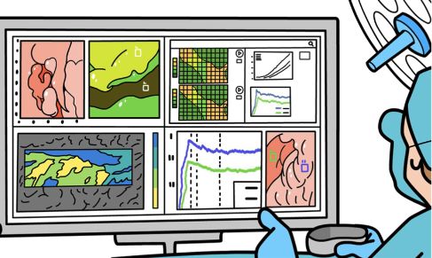
Introducing Al fluorescence guided surgery platform

UltraGreen dye icon

Enabled by fluorescence dye + data management

Data cloud icon

Using dynamic data capture, cloud based Al/ML applications

Surgical real time data icon

For real time surgical decision-making

illustration showing a surgeon getting real time data in operating room

Generating Patient Specific Data During Surgery Enabled by Fluorescence and Software

References:

1. H Vaassen, B Wermelink, S Manohar et al., Intraoperative quantification of fluorescence angiography for assessment of intestinal perfusion: in vivo exploration of clinical value; BJS Open. 2022 Jun; 6(3): zrac058; https://www.ncbi.nlm.nih.gov/pmc/articles/PMC9072211/
2. M Noltes, M Metman, W Heeman et al., A Novel and Generic Workflow of Indocyanine Green Perfusion Assessment Integrating Standardization and Quantification Toward Clinical Implementation; Observational Study Ann Surg. 2021 Dec 1;274(6):e659-e663. doi: 10.1097/SLA.0000000000004978; https://pubmed.ncbi.nlm.nih.gov/34145192/
3. C Lütken, M Achiam, J Osterkamp et al., Quantification of fluorescence angiography: Toward a reliable intraoperative assessment of tissue perfusion – A narrative review; Review Langenbecks Arch Surg. 2021 Mar;406(2):251-259. doi: 10.1007/s00423-020-01966-0. Epub 2020 Aug 21; https://pubmed.ncbi.nlm.nih.gov/32821959/